Dental X-rays often spark debate among patients and professionals alike. Many wonder if these images are truly necessary for maintaining oral health. This article explores the importance of dental X-rays in diagnosing issues that may not be visible during a regular examination and highlights when they’re essential for effective treatment. Understanding their role can help individuals make informed decisions about their dental care.
Understanding Dental X Rays
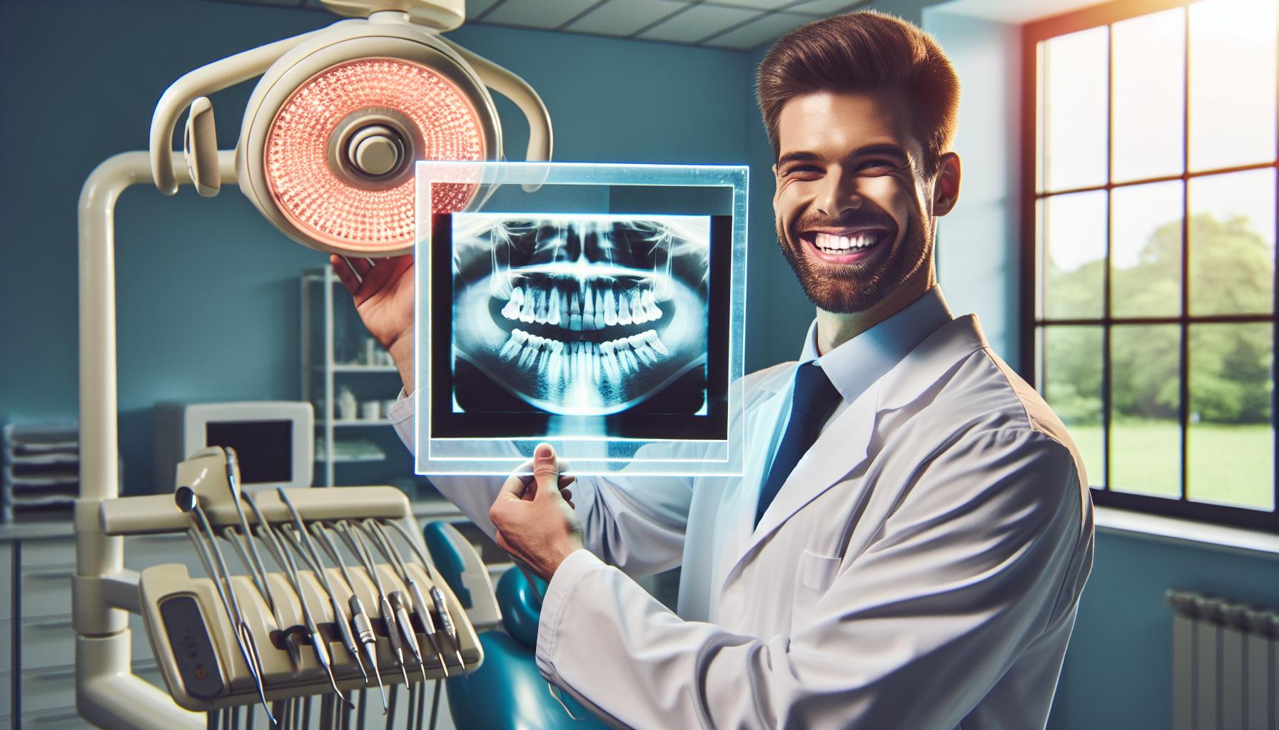
Dental X-rays play a crucial role in oral health by revealing issues not visible during a routine examination. These diagnostic tools help in identifying cavities, gum disease, and other dental conditions.
What Are Dental X Rays?
Dental X-rays are imaging tests that use a small amount of radiation to produce detailed pictures of teeth, gums, and surrounding bone structures. They assist dentists in diagnosing problems early, enabling timely interventions. Types of X-rays include intraoral and extraoral, each serving different diagnostic purposes.
Types of Dental X Rays
- Intraoral X-rays:
- Periapical X-rays: Capture the entire tooth, from crown to root, and show surrounding bone. Essential for diagnosing root problems and severe decay.
- Bitewing X-rays: Allow views of multiple teeth in one image, useful for detecting cavities between teeth and checking the level of bone support.
- Occlusal X-rays: Exhibit the position of teeth in the jaw and assist in assessing dental eruption and jaw abnormalities.
- Extraoral X-rays:
- Panoramic X-rays: Provide a broad view of the teeth and jaw in a single image. Helpful for evaluating the entire mouth and detecting impacted teeth.
- Cephalometric X-rays: Focus on the entire skull, aiding in orthodontic evaluations and treatment planning.
Understanding the types of dental X-rays and their functions allows patients to appreciate their necessity in comprehensive dental care.
The Importance of Dental X Rays

Dental X-rays play a crucial role in maintaining oral health by uncovering issues that may remain undetected during standard dental examinations.
Diagnosing Oral Health Issues
Dental X-rays provide detailed images of teeth, gums, and surrounding bone structures. Intraoral X-rays, including periapical and bitewing types, focus on specific areas, highlighting cavities, root issues, and bone loss. Extraoral X-rays, such as panoramic X-rays, give a broader view, aiding in detecting jaw problems and impacted teeth. These diagnostic tools allow dentists to form accurate treatment plans and address hidden conditions early, ultimately preventing more severe complications.
Preventive Care Benefits
Dental X-rays contribute significantly to preventive care by allowing for the early identification of potential problems. Regular X-ray examinations facilitate timely interventions, such as filling cavities before they worsen or managing gum disease effectively. By leveraging dental X-rays, patients can avoid extensive treatments, reduce costs, and ensure long-term oral health.
Common Concerns
Dental X-rays often raise questions regarding their necessity and safety. Understanding these concerns helps clarify their role in dental care.
Safety of Dental X-rays
Dental X-rays utilise minimal radiation exposure, with advancements in technology reducing risks significantly. Digital X-rays emit up to 80% less radiation than traditional film X-rays, making them safer for patients. The potential benefits of identifying dental issues early outweigh the risks associated with radiation exposure, particularly when appropriate safeguards are in place. Dentists always ensure that protective measures, such as lead aprons, are employed to shield patients from unnecessary radiation.
Frequency of Dental X-rays
The frequency of dental X-rays varies based on an individual’s oral health needs. For most patients, routine check-ups may require X-rays every 1 to 2 years. However, patients with specific dental issues may need more frequent imaging, allowing dentists to monitor conditions effectively. Factors influencing X-ray frequency include age, oral health history, and any existing dental problems. Regularly scheduled X-rays facilitate timely interventions, ensuring issues are addressed before they develop into more serious concerns.
Alternative Diagnostic Methods
Dental professionals employ several alternative diagnostic methods to assess oral health without relying solely on X-rays. These methods may offer additional perspectives on dental issues and help guide effective treatment. Diagnostic tools like the ProDENT Handheld X-Ray with scatter provide portable and efficient imaging, enabling dentists to perform accurate diagnostics even in challenging environments.
Clinical Examination Techniques
Clinical examination techniques involve thorough visual inspections and tactile assessments by dental professionals. During these examinations, dentists evaluate gums, tooth surfaces, and overall oral hygiene. They look for signs of decay, gum disease, and structural integrity. Dentists often use tools such as probes and mirrors to enhance visibility and access hard-to-reach areas. Certain symptoms, such as swelling or tenderness in the gums, may indicate underlying problems. Additionally, the use of specific diagnostic tests, such as vitality testing, can help assess tooth health without imaging.
Other Imaging Options
Other imaging options provide valuable insights into oral health when dental X-rays are unnecessary. For instance, Cone Beam Computed Tomography (CBCT) creates three-dimensional images of dental structures, offering a comprehensive view of teeth, bones, and soft tissues. This non-invasive option aids in precise diagnosis and treatment planning for complex cases. Alternatively, magnetic resonance imaging (MRI) can be used in particular situations, such as assessing soft tissue conditions. Ultrasound imaging may also assist in evaluating abscesses or cysts in soft tissues without the exposure to radiation associated with X-rays.
Conclusion
Dental X-rays play a pivotal role in maintaining oral health by uncovering hidden issues that might otherwise go unnoticed. They enable dentists to diagnose conditions early and develop effective treatment plans tailored to individual needs. While concerns about radiation exposure are valid, advancements in technology have significantly mitigated these risks.
Patients should recognise that the benefits of timely diagnosis and intervention often outweigh potential downsides. Understanding when and why dental X-rays are necessary empowers individuals to make informed decisions about their dental care. Ultimately, these diagnostic tools are essential for achieving and maintaining optimal oral health.
Frequently Asked Questions
What are the primary purposes of dental X-rays?
Dental X-rays are essential for diagnosing hidden dental problems such as cavities, gum disease, and root issues. They provide detailed images that help dentists create accurate treatment plans and allow for early detection of conditions that might not be visible during a standard examination.
How often should dental X-rays be taken?
The frequency of dental X-rays typically ranges from every 1 to 2 years for routine check-ups, depending on individual oral health needs. Patients with specific dental issues may require X-rays more frequently to monitor their condition effectively.
Are dental X-rays safe?
Yes, dental X-rays are safe. They use minimal radiation exposure, especially with advancements such as digital X-rays, which emit significantly less radiation than traditional film X-rays. Protective measures, like lead aprons, further reduce risks.
What types of dental X-rays are there?
There are two main types of dental X-rays: intraoral and extraoral. Intraoral X-rays include periapical, bitewing, and occlusal X-rays, providing detailed images of specific areas. Extraoral X-rays, such as panoramic and cephalometric X-rays, give broader views of the jaw and teeth.
What are the alternatives to dental X-rays?
Alternatives to dental X-rays include clinical examinations, vitality testing to assess tooth health, and advanced imaging techniques like Cone Beam Computed Tomography (CBCT) and magnetic resonance imaging (MRI) for complex cases. Ultrasound is also an option for evaluating soft tissue conditions.